Historically, there has been no safe, non-invasive and/or efficient method with deep penetration for modulating the human brain in vivo—stimulating or suppressing certain brain processes in a living person. Ultrasound, however, offers a promising approach, especially when used in combination with other non-invasive techniques.
Ultrasound—a decades-proven technique in which the echoes of high-frequency sound waves off internal body tissue are converted into sonogram images—can safely penetrate the human skull and be focused in different regions of the brain, probing responses. The penetration is on the order of several centimeters and can cover the entire brain. The weakness of the ultrasound approach is that the focus is on the order of 1cm, which is not as selective as some invasive techniques. In addition, the mechanism of probing the brain with ultrasound currently remains unknown, and control of the modulation method remains a challenge.
Still, ultrasound has been established as a safe modality that can induce thermal and mechanical effects for tapping into the underlying circuitry of the brain, and its strategic use in combination with other non-invasive approaches shows real potential for delivering breakthroughs for care of cancer, tumors, psychiatric, motor neural diseases, and a whole range of conditions via brain modulation. The next step is to marshaling more engagement and experimentation from across and beyond the neurology and biomedical engineering communities.
A mystery, still
The brain is a unique organ that dictates the function of the entire body, and, yet today, we still do not really understand how it works.
The technologies required to safely produce a complete, detailed mapping of the living human brain—all of the cells, all of the circuits—are not yet available. Resolution down to the cellular level (on the order of tens of microns) is required. While that capability is available via optical imaging, it is only within the head depth of a millimeter, so it reaches only the surface of the brain. To attain a deeper image, electrodes must be introduced. But doing so changes and can even damage the brain, undercutting the usefulness of invasive electrodes for understanding a healthy, human brain.
Current non-invasive techniques present their own tradeoffs: Modalities that deliver sufficient resolution are limited severely by the depth they can probe; other non-invasive methods that reach sufficient depth are limited in the image resolution they can deliver.
Understanding technological options
The ultrasound of the brain is not new. Medical ultrasound has been around since after World War II, and the brain was the number one organ that was imaged with ultrasound. Ultrasound waves can cross through the skull and give us some information about brain volume or more potential fluid in the brain, in the case of edema or from accidents.
But while ultrasound probes depths upwards to tens of centimeters—deep enough to map the entire brain—it fails to provide the cell-by-cell detail that is needed. Ultrasound’s focus on the order of a couple of millimeters delivers granularity no finer than at least a few hundred brain cells. Indeed, because of its limitations, ultrasound tends to be regarded as a kind of last resort for medical imaging. Other non-invasive techniques, however, also have their drawbacks in relation to work with the living human brain.
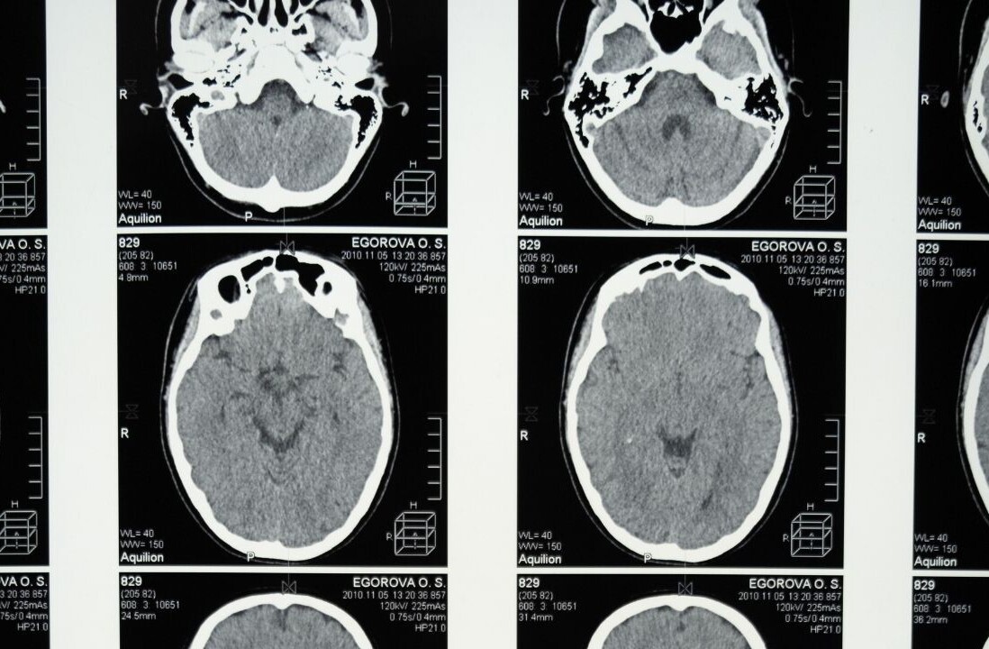
Computerized tomography (CT) scanning—in which multiple X-ray images are integrated to produce cross-section images of the internal body—is valuable but cannot be heavily relied upon for brain modulation in vivo. X-rays introduce ionizing radiation, so CT scans will damage the brain and other tissues around it if they are utilized too often.
Because it does not rely on X-rays, magnetic resonance imaging (MRI) is more conducive to brain modulation. Functional MRI processes (fMRI) are especially valuable for tapping into brain circuitry, looking at blood flow and exploring hemodynamic response. But, as far as probing the tissue, MRI is limited in its effectiveness.
Strategically combining methods
An approach that we are developing at Columbia University integrates ultrasound, MRI and electroencephalography (EEG), in which electrodes along with the scalp record electrical activity in the brain. Integrating EEG electrodes in the MRI is not straightforward because there are so many interferences and magnetic fields to be dealt with. But, by filtering out all this noise and getting an EEG to co-localize within fMRI response, we can basically probe the brain at the same time as with ultrasound. Waves are shined into the brain to mechanically or even thermally stimulate or inhibit a circuit.
Over the past decade, ultrasound through MRI guidance has been proven to be reproducible, safe enough, and reliable enough to be used in the living brain. The MRI shows the anatomical map to register the ultrasound transducer and the beam within the MRI system. In the United States, for example, this technique has been approved by the Food and Drug Administration (FDA) for non-invasive ablation of essential tremors. Within 20 minutes and without any incision, a person can be relieved of the nerve disorder and its uncontrollable shaking.
Frontiers of innovation
Today, instead of destroying a part of or burning somebody’s brain, we are exploring whether the same combination of technologies could be utilized to non-invasively modulate the brain. And mapping the whole structure of the brain is still another whole frontier.
What’s needed now is for more of the neurology and biomedical engineering communities to experiment with the technologies and these types of integrated approaches. Right now, unfortunately, it is not a plug-and-play scenario. We want manufacturers to potentially work with developing tools for neuroscientists, such as integrating into their MRI systems an ultrasound component.
More collaborative work needs to be done, but the promise to treat cancers, tumors, chronic pain, clinical depression, obsessive-compulsive disorder (OCD), Parkinson’s, alcoholism, comas, and a whole range of conditions by non-invasively stimulating or inhibiting function is tremendous.
This story is republished from TechTalks, the blog that explores how technology is solving problems… and creating new ones. Like them on Facebook here and follow them down here:
Get the TNW newsletter
Get the most important tech news in your inbox each week.


